40 simple microscope diagram with labels
Microscope Parts And Use Worksheet - Doeco 6 Best Images of Compound Microscope Parts Quiz Worksheet . Label the parts of the microscope. ... Microscope parts and use worksheet answer key along with labeling the parts of the microscope blank diagram available for. 8/7/2018 2 •simple •compound •stereoscopic. The best way to building a microscope parts and use worksheet. animal cell under electron microscope - Corrinne Simms Within the cell there is a shape of round with a circular structure of granulated part on the. Diagram Of Animal Cell Under Electron Microscope Labeled. As you can see in the above labeled plant cell diagram under light microscope there are 13 parts namely Cell membrane. These are both specific typesof cells.
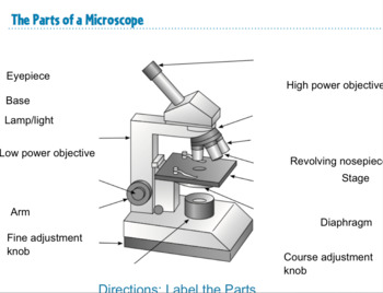
microscope parts and functions worksheet pdf - Alverta Overton This supports the body tube and makes a good handle for carrying the microscope. Microscope parts and functions worksheet answers. Body tube connects the eyepiece to the nosepiece usually moves during focusing Eyepiece - the lens you look through. Light passes from the objective lens to the eyepiece through this part of the microscope.

Simple microscope diagram with labels
Microscope Labeling - label microscope diagram, 32 picture of ... Microscope Labeling. Here are a number of highest rated Microscope Labeling pictures upon internet. We identified it from well-behaved source. Its submitted by supervision in the best field. We take on this nice of Microscope Labeling graphic could possibly be the most trending topic when we part it in google pro or facebook. Diagram Of Stomata Class 10 - Wiring Schematic Online Biology practicals for class 10 cbse. In order to complete the diagram of stomatal apparatus given below nuclei should be drawn in the parts marked. Delhi 2007c a a and b b a and c c b and c d a b and c. The epidermis is made up of single layer of cells. We can see stomata under the light microscope. Microscope Sketch - phycokey gloeocapsa images, micscape image gallery ... Microscope Sketch - 18 images - microscope drawing workshop flickr, microscope 4 coloring page free school coloring pages, premium vector hand drawn sketch of microscope in black, microscope sketch stock photography image 26513712,
Simple microscope diagram with labels. Cell The Label Diagram Plant Start studying Labeling a Plant Cell One of the distinctive aspects of a plant cell is the presence of a cell wall outside the cell membrane - Step 2: Draw a picture of your city Questions And Answers On Labeled/Unlebled Diagrams Of A Human Cell / Wa 6365 Simple Labeled Animal Cell Diagram Picture Unlabeled Plant Cell Free Diagram - Ii) give the main function of the parts labelled 5, 6, 7 ... microscope parts and functions worksheet pdf Some of the worksheets below are Parts and Function of a Microscope Worksheets with colorful charts and diagrams to help students familiarize with the parts of the microscope along with several important questions and activities with answers. _____ simple microscope a support entire microscope. microscope parts and functions pdf free download In the Word Bank check the box next to each part of the microscope after you have labeled it on the diagram. Built-in light sources An illuminator is built into the base of the. Click on the boxes to see the name and function of. Some of the worksheets below are Parts and Function of a Microscope Worksheets with colorful charts and diagrams to ... Simple Microscope - fibers free full text high strength and high ... Simple Microscope - 18 images - a microscope labeled micropedia, anatomy of microscope, types of microscopes and their uses science struck, microscopes lesson plans and lesson ideas brainpop educators,
Anatomy of the Epidermis with Pictures - Verywell Health Summary. The epidermis is composed of layers of skin cells called keratinocytes. Your skin has four layers of skin cells in the epidermis and an additional fifth layer in areas of thick skin. The four layers of cells, beginning at the bottom, are the stratum basale, stratum spinosum, stratum granulosum, and stratum corneum. Flagella: Structure, Arrangement, Function - Microbe Online Structure. The long helical filament of bacterial flagella is composed of many subunits of a single protein, flagellin, arranged in several intertwined chains. A flagellum consists of several components and moves by rotation, much like a propeller of a boat motor. The base of the flagellum is structurally different from the filament. PDF Color The Microscope Parts Key Answers Label And Color The Parts Of Both Microscopes - Ythoreccio Read PDF Color The Microscope Parts Key Answers obtained is the product of the eyepiece times that of the objective lens. You can easily switch objectives by turning the rotating nosepiece (E). Color the nosepiece blue-green . The coarse adjustment knob (B) is the larger on your microscope. animal cell mitosis color and label - ibg-world.com animal cell mitosis color and label; nike waffle one summit white 12 May 2022 class c water quality standards ...
polarizing microscope parts and functions pdf 872018 2 Simple Compound Stereoscopic Electron Simple Microscope Similar to a magnifying glass and has only one lens. Before exploring microscope parts and functions you should probably understand that the compound light microscope is more complicated than just a microscope with more than one lens. Some polarizing light microscopes have digital ... Using A Microscope Worksheet - math skills workshets Using A Microscope Worksheet. Using microscopes biology start practising. _____ simple microscope a) support entire microscope.Microscope Worksheet mustenjoy from mustenjoy.weebly.comCorrectly identify various parts of a brightfield microscope. The term microscope can be translated as "to view the tiny," because microscopes are used to study things that are too small to be easily observed ... PDF Parts Of A Microscope Answer Key Parts of a microscope with functions and labeled diagram Some of the basic parts of a microscope include: Eyepiece lens on top of the microscope and used to look through. Tube that connectes the ... microscope types, including simple, compound, and electron microscopes. Most microscopes used in a classroom Page 6/12. Download Free Parts Of A animal cell under electron microscope labelled - Cherly Pearce Animal Cell Diagram Under Microscope Labeled. ... Human cheek cells are made of simple squamous epithelial cells which are flat cells with a round visible nucleus that cover the inside lining of the cheekC. Living cells cannot be observed using an electron microscope because samples are placed in a vacuum. A typical animal cell is 1020 μm in ...
Microscope Labeling Worksheet 42 New Microscope Labeling Thinglink Worksheets Microscopes ...
Electron microscope - Wikipedia An electron microscope is a microscope that uses a beam of accelerated electrons as a source of illumination. As the wavelength of an electron can be up to 100,000 times shorter than that of visible light photons, electron microscopes have a higher resolving power than light microscopes and can reveal the structure of smaller objects. A scanning transmission electron microscope has achieved ...

Microscope Diagram Labeled, Unlabeled and Blank | Parts of a Microscope | Science printables ...
animal cell under microscope labeled - Rayford Runyon The diagram is very clear and labeled. Animal Cell Diagram Under Microscope Labeled. Animal cells are eukaryotic cells that contain a membrane-bound nucleus. A brief explanation of the different parts of an animal cell along with a. You see that many features are in common. Hair under a compound microscope.

A typical animal cell (as seen in an electron microscope) Medical Ima…
PDF Diagram For Labelling Parts Of Plant And Animal Cells Seen Circuit diagram - Simple circuits | Electricity and Circuits | Don't Memorise How to Draw Human Ear Diagram With Labelling Heat Class 7 Science - Thermometer - Clinical Thermometer - ... children can label the diagrams to reinforce their vocabulary on the topic.For teaching your KS1 (or Pre-Primary Age 3-5) you can use this lovely picture of ...

All Saints Online: Diagram for Labelling: Microscope
PDF Parts Of A Microscope Answer Key Parts of a microscope with functions and labeled diagram Some of the basic parts of a microscope include: Eyepiece lens on top of the microscope and used to look through. ... Today, there are a variety of microscope types, including simple, compound, and electron microscopes. Most microscopes used in a classroom setting are compound microscopes ...

White Blood Cells Diagram
PDF Well Label Diagram Of A Generalized Cell Label Diagram Of A Generalized Cellgetting this info. get the well label diagram of a generalized cell join that we find the money for here and check out the link. You could buy lead well label diagram of a generalized cell or get it as soon as feasible. You could quickly Page 2/41
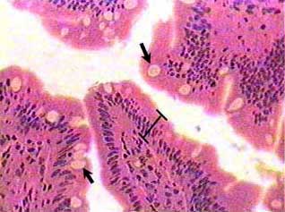
Simple columnar epithelium
animal cell under microscope labeled - Mad Thing Blogging Galleria Di ... Human cheek cells are made of simple squamous epithelial cells which are flat cells with a round visible nucleus that cover the inside lining of the cheekC. The animal cell diagram is widely. ... Animal Cell Diagram Under Microscope Labeled. Add a drop of purple stain specific for animals and cover with a cover slip. Draw a diagram of one cheek.

Diagram Of The Microscope - ClipArt Best
Light Microscope (Theory) - Amrita Vishwa Vidyapeetham Light microscope uses the properties of light to produce an enlarged image. It is the simplest type of microscope. Based on the simplicity of the microscope it may be categorized into: A) Simple microscope. B) Compound microscope. A) Simple microscope . It is uses only a single lens, e.g.: hand lens. Most of these are double convex or ...
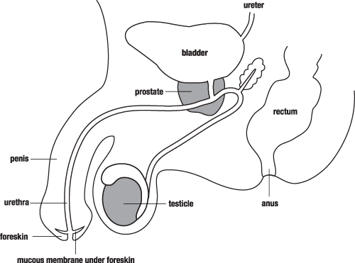
Male Reproductive System | Free Images at Clker.com - vector clip art online, royalty free ...
Microscope Sketch - phycokey gloeocapsa images, micscape image gallery ... Microscope Sketch - 18 images - microscope drawing workshop flickr, microscope 4 coloring page free school coloring pages, premium vector hand drawn sketch of microscope in black, microscope sketch stock photography image 26513712,
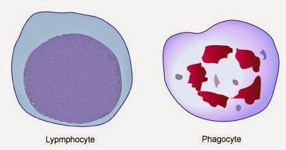
# 58 The immune system - Phagocytes | Biology Notes for A level
Diagram Of Stomata Class 10 - Wiring Schematic Online Biology practicals for class 10 cbse. In order to complete the diagram of stomatal apparatus given below nuclei should be drawn in the parts marked. Delhi 2007c a a and b b a and c c b and c d a b and c. The epidermis is made up of single layer of cells. We can see stomata under the light microscope.
All Saints Online: Diagram for Labelling: Microscope
Microscope Labeling - label microscope diagram, 32 picture of ... Microscope Labeling. Here are a number of highest rated Microscope Labeling pictures upon internet. We identified it from well-behaved source. Its submitted by supervision in the best field. We take on this nice of Microscope Labeling graphic could possibly be the most trending topic when we part it in google pro or facebook.
Simple Microscope Labeled Diagram - Micropedia

The Microscope: Create a Labelled Diagram | Teaching Resources

*Simple Squamous Epithelium*.........................(LOCATION: capillary walls, alveoli of ...

All Saints Online: Microscope Part Functions

The Microscope: Create a Labelled Diagram | Teaching Resources

Microscope Clip Art at Clker.com - vector clip art online, royalty free & public domain

anatomyforme: 2008-04-06
Post a Comment for "40 simple microscope diagram with labels"