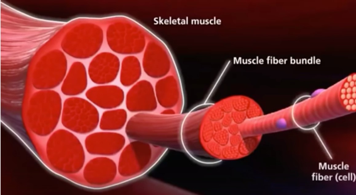
45 muscle fiber model with labels
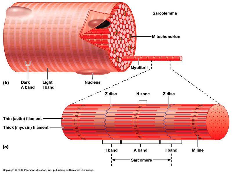
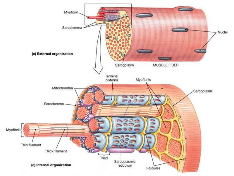
The Sarcomere and Sliding Filaments in Muscular Contraction: Definition ... Muscle fibers are composed of hundreds to thousands of contractile organelles called myofibrils. The myofibrils are packed tightly together in a parallel arrangement, much like Vienna sausages are... Label‐free 3D characterization of cardiac fibrosis in muscular ... Label-free 3D characterization of cardiac fibrosis in muscular dystrophy using SHG imaging of cleared tissue ... is a neuromuscular disease caused by mutations in the gene encoding dystrophin. It leads to repeated cycles of muscle fiber necrosis and regeneration and progressive replacement of fibers by fibrotic and adipose tissue, with ...
Parts of a Frog's Muscular System | Cuteness The muscle fibers occur in pairs and are attached to the skeleton by tendons. The pairing of the muscles is crucial to movement, as one serves to extend a limb, for example, while the other contracts it back again. Each striated muscle fiber in the frog's body is surrounded by a multilayered sarcolemma that is rich in adenosine triphosphate (ATP).
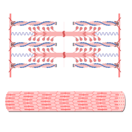
Muscle fiber model with labels
Anatomy, Head and Neck, Facial Muscles - NCBI Bookshelf The mimetic muscles are considered to be an extension of the superficial musculoaponeurotic system (SMAS) of the face, which is a fascial plane deep to the subcutaneous tissue but superficial to the muscles of mastication, running from the platysma in the neck up to the galea aponeurotica and the temporoparietal fascia under the scalp.[5] Alternative splicing diversifies the skeletal muscle transcriptome ... The size and fiber type composition of the gastrocnemius and quadriceps were differentially perturbed by prolonged spaceflight. On average, the CSA of muscle fibers composing the gastrocnemius decreased from 1170 ± 141 μm 2 to 856 ± 74 μm 2, representing an approximate 27% reduction (p < 0.01) in muscle fiber size as a result of extended exposure to microgravity (Fig. 2A, B). Chapter 8: Skeletal Muscle: Structure and Function - MHMedical.com Draw and label the microstructure of a skeletal muscle fiber. ... Compare and contrast the major biochemical and mechanical properties of the three primary types of muscle fibers found in human skeletal muscle. ... sliding filament model. slow-twitch fibers. summation. swinging lever-arm model. terminal cisternae. tetanus. transverse tubules.
Muscle fiber model with labels. Anatomy, Skeletal Muscle - StatPearls - NCBI Bookshelf The musculoskeletal system comprises one of the major tissue/organ systems in the body. The three main types of muscle tissue are skeletal, cardiac, and smooth muscle groups.[1][2][3] Skeletal muscle attaches to the bone by tendons, and together they produce all the movements of the body. The skeletal muscle fibers are crossed with a regular pattern of fine red and white lines, giving the ... (Get Answer) - Drag The Labels Onto The Diagram Of Muscle ... - Transtutors Drag The Labels Onto The Diagram Of Muscle Spindle Function. Reset Muscle Shortens Strelch Receptors In The Spinde Are Activated Throughout The Contraction Muscle Length Gamma Mator Neurons Activate Intrafusal Fibers Intrafusal Fibers Do Not Slacken So Firing Rate Remains Constant. Muscle Fiber Anatomy - artist spotlight laurie o keefe artists blogs ... Muscle Fiber Anatomy - 16 images - anatomy of a skeletal muscle fiber quiz, anatomy physiology review of skeletal muscle tissue youtube, muscle fiber anatomy definition, muscle fiber with motor end plate model physiology anatomy youtube, Muscles of Facial Expression | Anatomy | Geeky Medics The orbicularis oris muscle and modiolus act as the insertion site for this muscle. Action. Contraction of this muscle compresses the cheeks against the teeth (this action is especially useful in mastication and whistling). Innervation. The buccal branch of the facial nerve.¹; Blood supply. The buccal branch of the maxillary artery.¹
Muscular System - Muscles of the Human Body - Innerbody Muscular System. The muscular system is responsible for the movement of the human body. Attached to the bones of the skeletal system are about 700 named muscles that make up roughly half of a person's body weight. Each of these muscles is a discrete organ constructed of skeletal muscle tissue, blood vessels, tendons, and nerves. Neuromuscular Junction | Structure, Function, Summary & Clinical The neuromuscular junction is a chemical synapse between the motor neuron and the skeletal muscle fiber. It consists of a presynaptic terminal, synaptic cleft, and a postsynaptic membrane or cell. Presynaptic Terminal In case of neuromuscular junction, the presynaptic terminal is an axonal terminal of a motor neuron. Skeletal Muscle Slide Labeled - can you name the structures of a ... Skeletal Muscle Slide Labeled - 16 images - how the skeletal system works with the muscular system human body systems, skeletal muscle anatomical features, chapter 7 page 8 histologyolm 4 0, skeletal muscle structure proprofs quiz, Muscle forces and fascicle behavior during three hamstring exercises ... We hypothesize that the NHC shows the highest peak hamstring muscle forces due to the potential eccentric muscle fiber action 29 and hypothesize that the DL shows the largest excursion in biceps femoris fascicle length ... Raw marker data were labeled in Vicon Nexus, gaps were filled with a combination of spline and rigid body fills and ...
Muscles of the Posterior Forearm | Anatomy | Geeky Medics Figure 4.Brachioradialis muscle. 2 Extensor carpi radialis longus. The extensor carpi radialis longus has a relatively short muscle belly and longer tendon. It is this long tendon, and its superficial appearance, that identifies it from the extensor carpi radialis brevis.. Function: wrist extension and abduction; Origin: lateral supracondylar ridge and lateral epicondyle The Muscles of Facial Expression - Orbital Group - TeachMeAnatomy The oral group of muscles consists of the orbicularis oris, buccinator, and various smaller muscles. Orbicularis Oris The fibres of the orbicularis oris enclose the opening to the oral cavity. Attachments: Arises from the maxilla and from the other muscles of the cheek. It inserts into the skin and mucous membranes of the lips. Anatomical Models and Keys: Anatomy Models - NEOMED Saunders Heart Model . Female Pelvis on Stand. Denoyer Geppert Heart. Kidney Microanatomy . Stomach. Muscle Fiber. Bone Structure. Liver Microanatomy . Heart and Diaphragm . Larynx . Base of Head . Small Liver Model . Liver Denoyer Model . Kidney Denoyer Model Disc/MRI Head . Smooth Muscle . Heart and Lung Larynx is missing . Brain Stem ... Fluorescent labeling of abundant reactive entities (FLARE) for ... - Nature The cross-striations of skeletal muscle fibers are well visualized in c and d, and fibrocytes and erythrocytes are distinctively labeled in d. Zoom-in views of the breast core from the boxed region...
Muscle Physiology • An Introduction
Type IIa Muscle Fibers: Training for Explosiveness | ISSA Type IIx Muscle Fibers There is a second type of fast twitch muscle fiber called type IIx. These are even faster and more powerful than type IIa. They are also even more inefficient, fatiguing very quickly. Type IIx fibers are used for activities of very short duration that require significant power and strength.
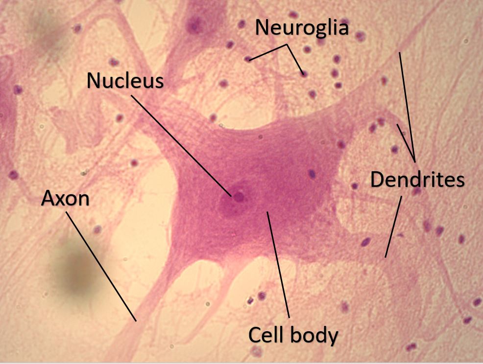
21 Fresh Neuron Label And Function
Muscle anatomy reference charts: Free PDF download | Kenhub This muscle chart eBook covers the following regions: Inner hip & gluteal muscles Anterior, medical and posterior thigh muscles Anterior, lateral and posterior leg muscles Dorsal and plantar foot muscles This eBook contains high-quality illustrations and validated information about each muscle. It is available for free. Download free PDF (8.5MB)

V Ling: 08.10
Free Worksheets for the Muscular System - Homeschool Giveaways Download free printables here and follow instructions to make your own lapbook to show off everything you know now about how the muscular system works. Inside Out Anatomy: Muscles Worksheet - Download a muscular system worksheet set to explore all the different types of muscles and how they help us move. Muscle Labeling Worksheet - Label ...

Muscle Fiber Types
Skeletal Muscle Fiber Definition and Anatomy - Study.com Type 1 muscle fibers are often found in muscles that must work for long periods of time, like those involved in maintaining posture or walking. Type 2A and 2B muscle fibers are fast-twitch fibers,...

Ch10 Skeletal Muscle Fiber IMAGES Flashcards | Quizlet
Drag the correct label to the appropriate location to identify the ... -Labeling Activity: Sarcoplasmic Reticulum And T Tubules In The Skeletal Muscle Fiber Drag The Correct Label To The Appropriate Location To Identify The Parts Of The Skeletal Muscle Fiber. Reset Help Mitochondria Part Of A Skeletal Muscle Fiber... Q: View Answer Q: Posted one year ago Q: Posted 4 months ago Q: Posted 3 months ago

Muscle Fiber Types | Renaissance Man Journal
Targeting necroptosis in muscle fibers ameliorates ... - Nature The severity of myositis was graded histologically on the scales of 1-6, where 1 = involvement of 1 muscle fiber, 2 = involvement of 2-5 muscle fibers, 3 = involvement of 6-15 muscle fibers ...
Microscopic Muscle Anatomy and Muscle Physiology week #10 Flashcards ...
Back Muscles: Anatomy, Function, Treatment - Verywell Health Your back consists of a complex array of bones, discs, nerves, joints, and muscles. The muscles of your back support your spine, attach your pelvis and shoulders to your trunk, and provide mobility and stability to your trunk and spine. The anatomy of your back muscles can be complex. There are several different layers of muscles in your back ...

10.06.20 - Types of Muscle Fibers - Amanda Jackson Whitney | Library ...
Ultrastructure of Muscle - Skeletal - Sliding Filament - TeachMeAnatomy The sliding filament model describes the mechanism of skeletal muscle contraction Actin and Myosin Muscle fibres are formed from two contractile proteins - actin and myosin. Myosin filaments have many heads, which can bind to sites on the actin filament. Actin filaments are associated with two other regulatory proteins, troponin and tropomyosin.
Anatomy Exam 2 Flashcards | Easy Notecards
Learn all muscles with quizzes and labeled diagrams | Kenhub Labeled diagram View the muscles of the upper and lower extremity in the diagrams below. Use the location, shape and surrounding structures to help you memorize each muscle. Once you're feeling confident, it's time to test yourself. Unlabeled diagram See if you can label the muscles yourself on the worksheet available for download below.

Password Needed! | Quizlet
kb291354's Favorite Games muscle fiber model. Medicine. Played 0 times Faved 3 weeks ago. Muscle Fiber. Science. Played 7,693 times Faved 3 weeks ago. Label the Ethmoid. Medicine. Played 1,265 times Faved 3 weeks ago. Bones and Bony Landmarks of the Vertebral Column. Science Editor's Choice. Played 20,564 times ...
Plant Cell Model Cake – Eclectic Homeschooling
Plus, How to Make the Most of Leg Exercises - Shape When it comes to the front of your legs, there are two muscle groups — the anterior upper leg muscles (i.e. your thigh) and the anterior lower leg muscles (i.e. your shin). There are four parts of your quadriceps: rectus femoris, vastus lateralis, vastus medialis, and vastus intermedius. The tibialis anterior is the strip of muscle that makes ...
272: FREE Training Series: Everything You Need To Know About Building ...
Chapter 8: Skeletal Muscle: Structure and Function - MHMedical.com Draw and label the microstructure of a skeletal muscle fiber. ... Compare and contrast the major biochemical and mechanical properties of the three primary types of muscle fibers found in human skeletal muscle. ... sliding filament model. slow-twitch fibers. summation. swinging lever-arm model. terminal cisternae. tetanus. transverse tubules.
MUSCULAR SYSTEM ANATOMY:Muscle fiber with sarcomere model description ...
Alternative splicing diversifies the skeletal muscle transcriptome ... The size and fiber type composition of the gastrocnemius and quadriceps were differentially perturbed by prolonged spaceflight. On average, the CSA of muscle fibers composing the gastrocnemius decreased from 1170 ± 141 μm 2 to 856 ± 74 μm 2, representing an approximate 27% reduction (p < 0.01) in muscle fiber size as a result of extended exposure to microgravity (Fig. 2A, B).
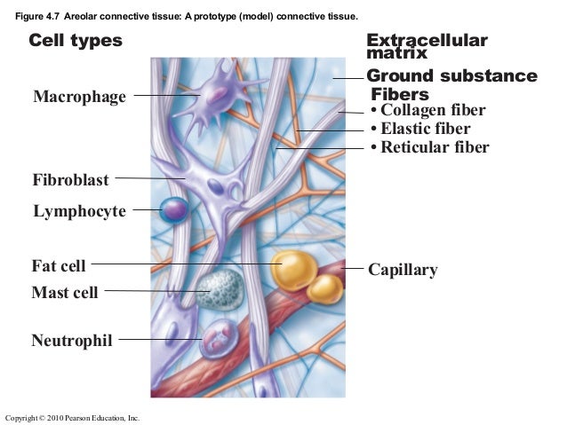
Ex 6a connective_tissues
Anatomy, Head and Neck, Facial Muscles - NCBI Bookshelf The mimetic muscles are considered to be an extension of the superficial musculoaponeurotic system (SMAS) of the face, which is a fascial plane deep to the subcutaneous tissue but superficial to the muscles of mastication, running from the platysma in the neck up to the galea aponeurotica and the temporoparietal fascia under the scalp.[5]

3 Types of Muscle Fiber in your Body | #ScienceSaturday

32 Label The Structures Of A Skeletal Muscle Fiber - Label Design Ideas ...

Skeletal Muscle Cell Model | A&P.2.Skin.Bone.Muscle | Pinterest ...
Post a Comment for "45 muscle fiber model with labels"