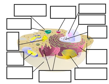
42 cell diagram and labels
Animal Cells: Labelled Diagram, Definitions, and Structure - Research Tweet The endoplasmic reticulum (s) are organelles that create a network of membranes that transport substances around the cell. They have phospholipid bilayers. There are two types of ER: the rough ER, and the smooth ER. The rough endoplasmic reticulum is rough because it has ribosomes (which is explained below) attached to it. Free Cell Diagram Software with Free Templates - EdrawMax - Edrawsoft An animal cell diagram describes a cell structure enclosed by a plasma member, and it has a nucleus with a membrane and organelles. Neuron Diagram A neuron diagram describes the three parts of a Neuron: dendrites, an axon, a cell body, or soma. Cell Membrane Diagram
Labeling a Cell Diagram | Quizlet Cell Wall This gives shape and support to the plant cell. It surrounds the cell and protects the other parts of the cell. Chloroplasts This is where the plant cell's chlorophyll is stored. This is what the plant uses to make its own food (photosynthesis). This is also what makes plant cells have a green-like color. Plant cells Are circular in shape
Cell diagram and labels
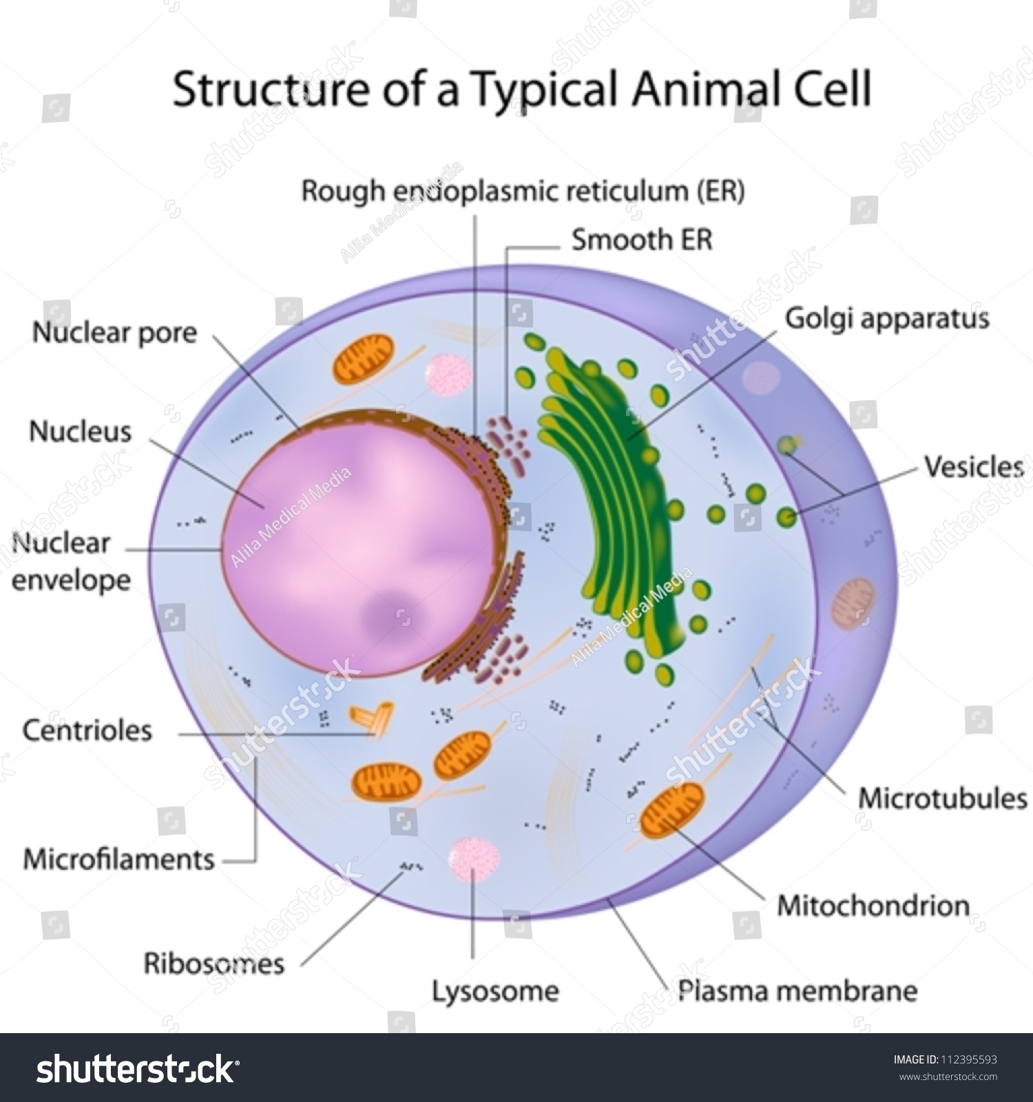
Plant Cell - Definition, Structure, Function, Diagram & Types - BYJUS The primary function of the cell wall is to protect and provide structural support to the cell. The plant cell wall is also involved in protecting the cell against mechanical stress and providing form and structure to the cell. It also filters the molecules passing in and out of it. The formation of the cell wall is guided by microtubules. Eukaryotic Cell Labeled Diagram | Quizlet Plasma membrane 9. phospholipid bilayer with embedded proteins that selectively regulates the flow of materials into and out of the cell. Lysosome 10. Contains enzymes for intracellular digestion of macromolecules and of organelles, the latter during the normal process of organelle replacement. type of cell. Learn the parts of a cell with diagrams and cell quizzes Cell diagram unlabeled It's time to label the cell yourself! As you fill in the cell structure worksheet, remember the functions of each part of the cell that you learned in the video. Doing this will help you to remember where each part is located. Click the links below to download the labeled and unlabeled eukaryotic cell diagrams.
Cell diagram and labels. PDF Human Cell Diagram, Parts, Pictures, Structure and Functions One of the few cells in the human body that lacks almost all organelles are the red blood cells. The main organelles are as follows : cell membrane endoplasmic reticulum Golgi apparatus lysosomes mitochondria nucleus perioxisomes microfilaments and microtubules 2 Animal and Plant Cell Worksheets - Super Teacher Worksheets Plant Cell Parts (Color Poster) FREE. This is a basic illustration of a plant cell with major parts labeled. Labels include nucleus, chloroplast, cytoplasm, membrane, cell wall, and vacuole, and mitochondrion. Use it as a poster in your classroom or have students glue it into their science notebooks. View PDF. Cell diagram with labels - Graph Diagram This human anatomy diagram with labels depicts and explains the details and or parts of the Cell Diagram With Labels.Human anatomy diagrams and charts show internal organs, body systems, cells, conditions, sickness and symptoms information and/or tips to ensure one lives in good health. 03 Label the Cell Diagram | Quizlet Start studying 03 Label the Cell. Learn vocabulary, terms, and more with flashcards, games, and other study tools. ... cell diagram. 18 terms. lugo_janet. Sets found in the same folder. 03 Organelle Functions. 14 terms. muskopf1. 07 Cell Labeling. 11 terms. muskopf1. 03 Cell Transport. 12 terms.
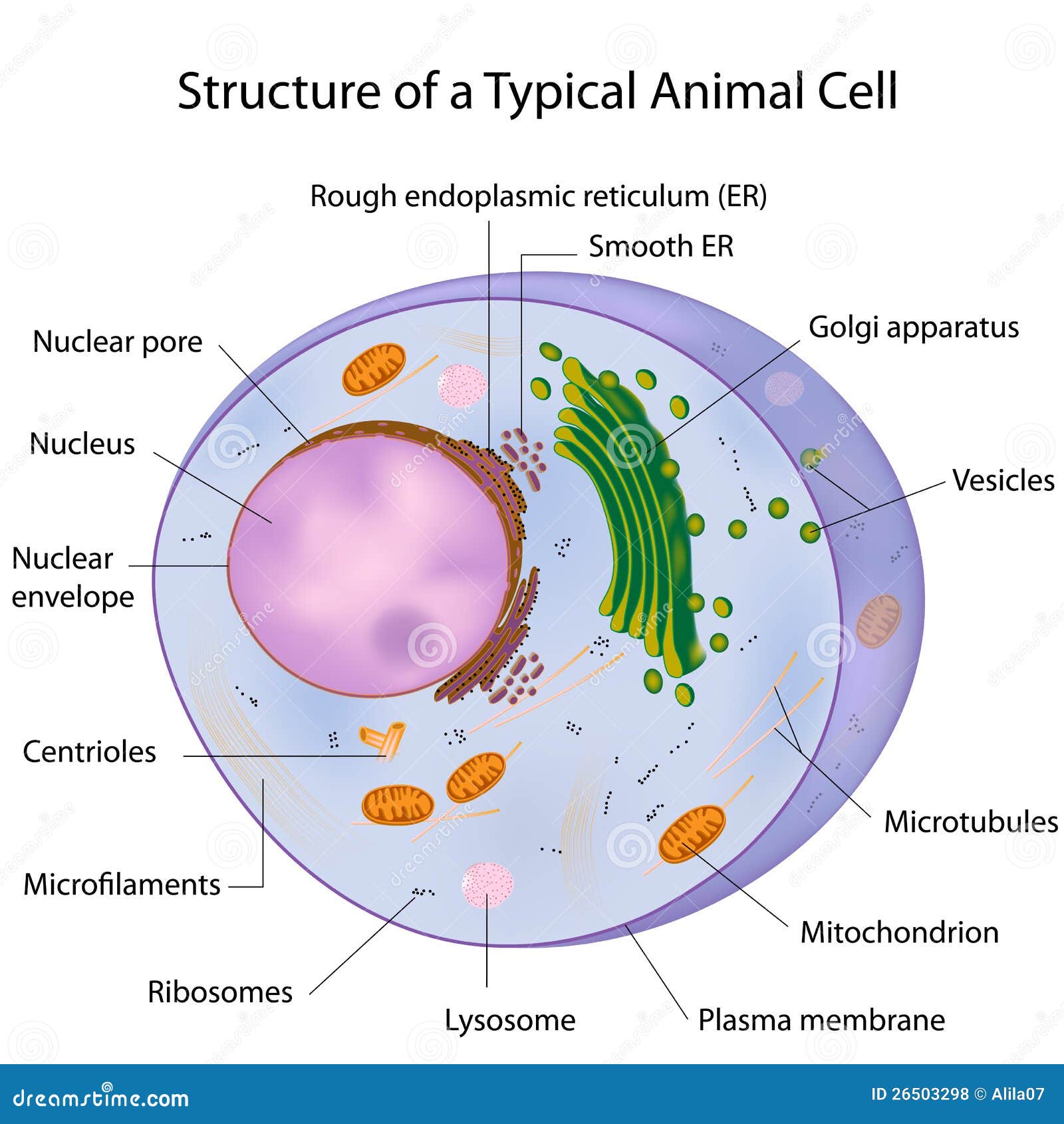
Animal Cell - Structure, Function, Diagram and Types - BYJUS The cell is the structural and functional unit of life. These cells differ in their shapes, sizes and their structure as they have to fulfil specific functions. Plant cells and animal cells share some common features as both are eukaryotic cells. However, they differ as animals need to adapt to a more active and non-sedentary lifestyle. Cell: Structure and Functions (With Diagram) - Biology Discussion Eukaryotic Cells: 1. Eukaryotes are sophisticated cells with a well defined nucleus and cell organelles. 2. The cells are comparatively larger in size (10-100 μm). 3. Unicellular to multicellular in nature and evolved ~1 billion years ago. 4. The cell membrane is semipermeable and flexible. 5. These cells reproduce both asexually and sexually. A Well-labelled Diagram Of Animal Cell With Explanation - BYJUS The animal cell diagram is widely asked in Class 10 and 12 examinations and is beneficial to understand the structure and functions of an animal. A brief explanation of the different parts of an animal cell along with a well-labelled diagram is mentioned below for reference. Also Read Different between Plant Cell and Animal Cell Structure of Cell: Definition, Types, Diagram, Functions - Embibe Cells are the fundamental structural and functional unit of all living beings including plants, animals and microorganisms. All living organisms in this universe are made up of cells. We cannot see cells with naked eyes as they are only \(10\) microns in size whereas human eyes cannot see objects less than \(100\) microns.
CELL MEMBRANE LABEL Diagram | Quizlet identifies or labels the cell. Receptor protein. receives information. Heads. part of the phospholipid that loves water (hydrophili) - points to the most outside and inside of cell. Tails. part of phospholipid that hates water (hydrophobic); points to the interior or Inside. Phospholipid Bilayer. 2 layers of fat - tails point in toward each ... Cell Diagram To Label Teaching Resources | Teachers Pay Teachers Cells: Cell Cycle (Mitosis) Diagram to Label by Lori Maldonado 5.0 (10) $2.00 PDF Students will label each of the stages of the cell cycle (mitosis). Plant Cell: Diagram, Types and Functions - Embibe Exams Q.2. How to make a model of a plant cell diagram step by step procedure? Ans: The plant cell diagram can be checked above and on a similar pattern the diagram can be created. Q.3. Why do plant cells possess large-sized vacuoles? Ans: Vacuole functions in the storage of substances, maintenance of osmolarity and sustaining turgor pressure. Q.4. Labeled Plant Cell With Diagrams | Science Trends The parts of a plant cell include the cell wall, the cell membrane, the cytoskeleton or cytoplasm, the nucleus, the Golgi body, the mitochondria, the peroxisome's, the vacuoles, ribosomes, and the endoplasmic reticulum. Parts Of A Plant Cell The Cell Wall Let's start from the outside and work our way inwards.
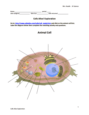
Blank Animal Cell Diagram To Label
Cell Organelles- Definition, Structure, Functions, Diagram Cilia and Flagella are tiny hair-like projections from the cell made of microtubules and covered by the plasma membrane. Structure of Cilia and Flagella Cilia are hair-like projections that have a 9+2 arrangement of microtubules with a radial pattern of 9 outer microtubule doublet that surrounds two singlet microtubules.
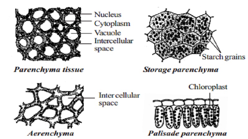
Permanent tissue: characteristics, types and functions - Online Biology Notes
A Labeled Diagram of the Plant Cell and Functions of its Organelles ... A Labeled Diagram of the Plant Cell and Functions of its Organelles We are aware that all life stems from a single cell, and that the cell is the most basic unit of all living organisms. The cell being the smallest unit of life, is akin to a tiny room which houses several organs. Here, let's study the plant cell in detail...
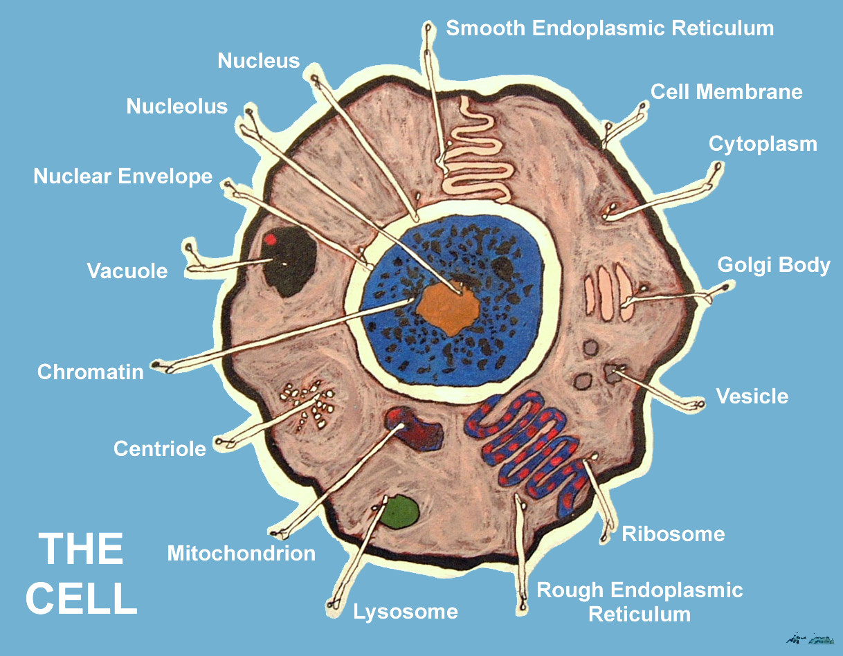
A Typical Cell, Labeled Royalty Free Stock Photos - Image: 26503298
Learn the parts of a cell with diagrams and cell quizzes Cell diagram unlabeled It's time to label the cell yourself! As you fill in the cell structure worksheet, remember the functions of each part of the cell that you learned in the video. Doing this will help you to remember where each part is located. Click the links below to download the labeled and unlabeled eukaryotic cell diagrams.

This is the Cell Wall. It protects the plant cell from an...
Eukaryotic Cell Labeled Diagram | Quizlet Plasma membrane 9. phospholipid bilayer with embedded proteins that selectively regulates the flow of materials into and out of the cell. Lysosome 10. Contains enzymes for intracellular digestion of macromolecules and of organelles, the latter during the normal process of organelle replacement. type of cell.
Typical Cell Labeled Stock Illustration 112395266 - Shutterstock
Plant Cell - Definition, Structure, Function, Diagram & Types - BYJUS The primary function of the cell wall is to protect and provide structural support to the cell. The plant cell wall is also involved in protecting the cell against mechanical stress and providing form and structure to the cell. It also filters the molecules passing in and out of it. The formation of the cell wall is guided by microtubules.
TIGER - NCSSM Distance Education and Extended Programs
Cell Diagram To Label - Pensandpieces
A Typical Cell, Labeled Stock Vector Illustration 112395593 : Shutterstock

Mr. Reynolds Science Jarrell Intermediate School: May 2014
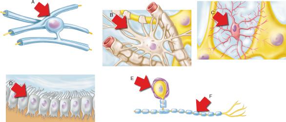
Print A&P Chapter 11 Fundamentals of the Nervous System and Nervous Tissue flashcards | Easy ...

Cell Diagram To Label - Pensandpieces
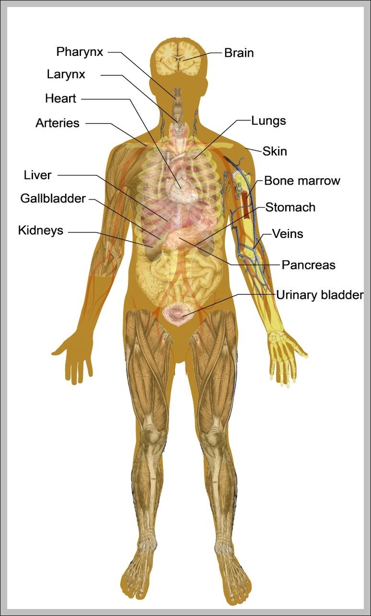
anatomy of the male body 744×1293 | Anatomy System - Human Body Anatomy diagram and chart images

The human egg cell explained for egg donors | Altrui

Human digestive system diagram & function explained
Post a Comment for "42 cell diagram and labels"