42 ear anatomy without labels
255 Human Ear Diagram Premium High Res Photos - Getty Images external auditory canal of human ear (with labels). - human ear diagram stock illustrations engraved antique, anatomy of the ear and nose engraving antique illustration, published 1851 - human ear diagram stock illustrations Ear Diagram and Labeling Worksheet / Worksheet - Twinkl This allows you to tailor the task to the individual abilities of your learners. The first worksheet presents an ear with annotations showing the first letters of its key features. For example, a label marked 'P' links to the Pinna (outer ear). The second page shows an ear diagram without labels. The final page shows the labels linking to the ...
Ear Anatomy - Outer Ear | McGovern Medical School The outer ear is made up of cartilage and skin. There are three different parts to the outer ear; the tragus, helix and the lobule. EAR CANAL The ear canal starts at the outer ear and ends at the ear drum. The canal is approximately an inch in length. The skin of the ear canal is very sensitive to pain and pressure.

Ear anatomy without labels
Male anatomy drawing labeled Male Anatomy Stock Photos And Images. 3d illustration, hip painful skeleton x-ray, medical concept. The legs of a man. sports legs men. realistic feet of a man. vector illustration. Human anatomy layout of internal organs in male body. isolated on black background. vector illustration. 3d rendering, conceptual human muscle, skin shade anatomy. 2022.7. 1. The Ear: Anatomy, Function, and Treatment - Verywell Health Anatomy The ear is divided into three portions: the outer ear, the middle ear, and the inner ear. Outer Ear The outer ear includes the visible outer portion of the ear and the ear canal. 2 Auricle: The outwardly visible part of the ear is composed of skin and cartilage, and attaches to the skull. Anatomical Line Drawings - Medscape Ear, Nose & Throat; Endocrine; Full Body; Gastrointestinal; Lymphatic; Musculoskeletal; ... go to drawing with labels go to drawing without labels; Surface Anatomy - anterior view & posterior view ...
Ear anatomy without labels. Anatomy, medical imaging and e-learning for healthcare IMAIOS and selected third parties, use cookies or similar technologies, in particular for audience measurement. Cookies allow us to analyze and store information such as the characteristics of your device as well as certain personal data (e.g., IP addresses, navigation, usage or geolocation data, unique identifiers). Cefdinir Antibiotic Side Effects, Uses (Strep, Middle Ear) & Dosage Cefdinir is an antibiotic in the cephalosporin drug class prescribed to treat infections, for example, middle ear, tonsillitis, strep throat, bronchitis, and sinusitis. Common side effects are nausea, abdominal pain, loose stools, and vaginitis. Dosage and pregnancy and breastfeeding safety information are included. Amazon.com: Flents Foam Ear Plugs, 10 Pair with Case for … If you have average ear canals and want the most noise blocking ear plugs, I would recommend these.Light Green Flents Protech Contour:I like contour ear plugs because my ears are sensitive. These are pretty large ear plugs and not the best for those with small ears. They were the best price of all the earplugs I tried. If you have average-larger ear canals, are a side sleeper, or … Anatomy coloring books: How to use & free PDF | Kenhub Sep 30, 2021 · Generally, an anatomy coloring book will divide subject matter into sections, with each section containing many topics. For each topic you will find black and white anatomical drawings, often accompanied by labels, related text and terminology. Tired of keeping track of so many study materials?
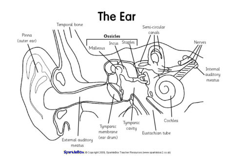
Human Body Parts Images Without Labels - Free Vector Download 2020 Human ear diagram with labels and label of anatomy labeling the ear purposegames nose diagram with label diagrams all labels human ear the ear diagram without labels anatomy human charts. Illustration Of Body Parts Labels It is certainly the most widely studied structure the world over. Human body parts images without labels. Download body ... Human Ear Anatomy - Parts of Ear Structure, Diagram and Ear Problems The external (outer) ear consists of the auricle, external auditory canal, and eardrum (Figure 1 and 2). The auricle or pinna is a flap of elastic cartilage shaped like the flared end of a trumpet and covered by skin. The rim of the auricle is the helix; the inferior portion is the lobule. Ligaments and muscles attach the auricle to the head. Anatomy of the Ear | Inner Ear | Middle Ear | Outer Ear auditory canal (also called the ear canal) eardrum outer layer (also called the tympanic membrane) The outer part of the ear collects sound. Sound travels through the auricle and the auditory canal, a short tube that ends at the eardrum. The Middle Ear The middle ear includes: eardrum cavity (also called the tympanic cavity) Human Ear Diagram - Bodytomy External Auditory Canal: External auditory canal or ear canal, is the channel from which the sound enters from the outside ear to the eardrum. Eardrum/Tympanic Membrane: It is the thin membrane located between the external ear canal and the middle ear. Cochlea: Cochlea is tiny conical structure situated in the inner ear that resembles a snail shell. It is responsible for converting sound vibrations into nerve signals that are sent to the brain.
Human penis - Wikipedia The human penis is an external male intromittent organ that additionally serves as the urinary duct.The main parts are the root (radix); the body (corpus); and the epithelium of the penis including the shaft skin and the foreskin (prepuce) covering the glans penis.The body of the penis is made up of three columns of tissue: two corpora cavernosa on the dorsal side and corpus … Human Middle Ear Anatomy Cross Section View With Labels Stock Photo ... Description Computer generated image of the human middle ear bones and inner ear with anatomical labeling. 1 credit Essentials collection for this image $4 with a 1-month subscription (10 Essentials images for $40) Continue with purchase View plans and pricing Includes our standard license. Add an extended license. Credit: Hank Grebe Ear Anatomy without Labels, Digital Art - Shutterstock Ear Anatomy Without Labels Digital Art Stock Illustration 530108302 Download for free See more Popularity score High Usage score High usage Superstar Shutterstock customers love this asset! Item ID: 530108302 Ear Anatomy without Labels, Digital Art Formats 8976 × 6201 pixels • 29.9 × 20.7 in • DPI 300 • JPG Human Ear Diagram Without Labels - ear anatomy diagram, ear diseases ... Human Ear Diagram Without Labels - 18 images - human ear diagram with label health images reference, blank digestive system no labels news word, ear anatomy poster laminated 24x16 anatomical new chart ebay, hearing loss resources for phoenicians,
Label the Ear Worksheets (SB6635) - SparkleBox
Image result for ear structure without label - Pinterest Human Ear Anatomy Image result for ear structure without label P Passant Ghonim 20 followers More information Image result for ear structure without label Find this Pin and more on • S C I E N C E • by Passant Ghonim. Human Ear Anatomy Human Anatomy Picture Label Templates Microsoft Word Templates Skeletal System Games Human Ear Diagram

This excellent ear diagram labels all the important parts of the human ear system. The labeled ...
Ear (Anatomy): Overview, Parts and Functions | Biology Dictionary The human ear picks up and interprets high-frequency vibrations of air, while the sound-sensing organs of aquatic animals are designed to pick up high-frequency vibrations in water. Most vertebrates have two ears: one on either side of the head. In some animals, including most mammals, the ear is also used for balance.

Oral Cavity Gallery - Medical Information Illustrated
EnchantedLearning.com | Worksheets, Activities, Crafts & More Moved Permanently. The document has moved here.

Medical Science
Ear Labels Flashcards | Quizlet Terms in this set (16) auricle external auditory canal tympanic membrane malleus (hammer) Incus (anvil) stapes (stirrup) auditory/eustachian/pharyngotympanic tube vestibules semicicular canals Ampulla of semicircular canals round window oval window and round window cochlea snail cochlear duct in cochlea vestibular nerve
Anatomy of ear
Best 10 Anatomy Apps - Last Updated July 26, 2022 - AppGrooves May 01, 2018 · This app is absolutely phenomenal. I have used it for 6 years now. The fact that you are able to explore in zoomable detail the thousands of muscles, tendons, ligaments joints, bones, organs, attachment points, connections, surfaces, etc., has been so enlightening for me in exploring the anatomy involved in the random injuries I have passed in life, including injuries that I never knew of ...
Ear | ClipArt ETC
14,026 Human ear anatomy Images, Stock Photos & Vectors - Shutterstock Human ear anatomy royalty-free images 14,026 human ear anatomy stock photos, vectors, and illustrations are available royalty-free. See human ear anatomy stock video clips Image type Orientation Color People Artists Sort by Popular Healthcare and Medical Anatomy ear cochlea medicine inner ear organ human body biology diagram Next of 141

Susan Price's 'Nennius' Blog: I Want To Write!
Enchanted Learning Moved Permanently. The document has moved here.

Pin by elizabeth wright on Free time | Human skull anatomy, Skull anatomy, Skull
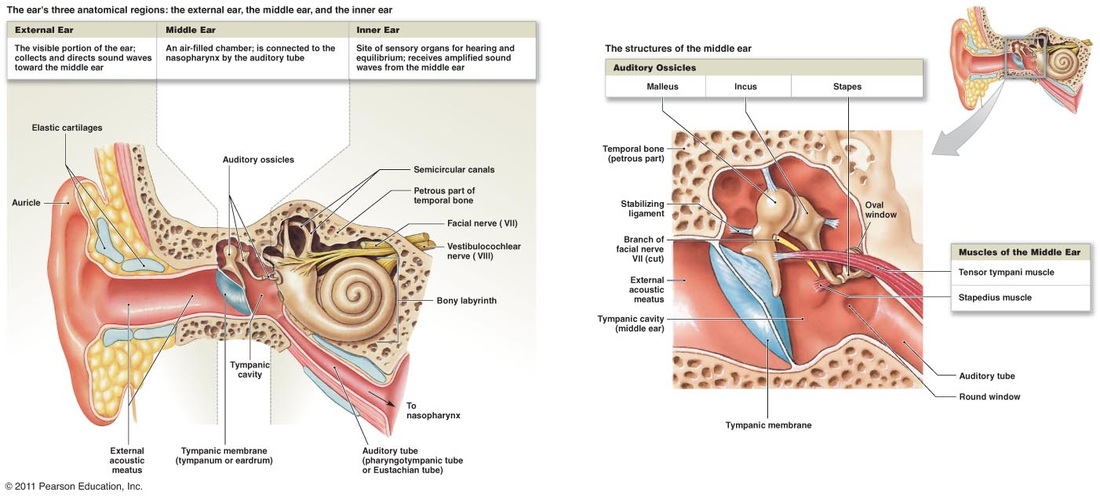
Anatomy of the Ear | Geeky Medics The middle ear, also referred to as the tympanic cavity, is an air-filled section in the temporal bone and is lined with a mucous membrane. 1,2,3 It can be split into two main sections: The tympanic cavity proper: this is the space directly internal to the tympanic membrane The epitympanic recess: this is the space behind pars flaccida
(184).jpg)
Anatomy Of The Ear - ProProfs Quiz
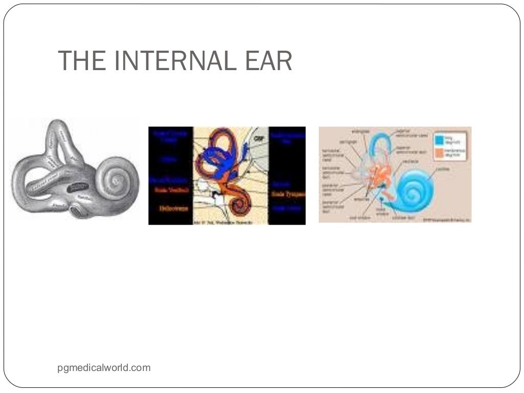
Well-Labelled Diagram Of Ear With Explanation - BYJUS Eustachian Tube is a tube that connects the middle ear to the back of the nose. It helps to maintain equal pressure in the middle ear which facilitates the proper transmission of sound waves. The Inner ear consists of Cochlea that comprises the nerves of hearing. Semicircular canals contain the receptors that help in maintaining balance.

Category:SVG all portions of the human ear - Wikimedia Commons
Normal chest MDCT with anatomic labels | e-Anatomy - e-Anatomy … Mar 10, 2022 · IMAIOS and selected third parties, use cookies or similar technologies, in particular for audience measurement. Cookies allow us to analyze and store information such as the characteristics of your device as well as certain personal data (e.g., IP addresses, navigation, usage or geolocation data, unique identifiers).

32 Label The Ear - Labels 2021
Anatomy of the eye: Quizzes and diagrams | Kenhub How to learn the parts of the eye. Found within two cavities in the skull known as the orbits, the eyes are surrounded by several supporting structures including muscles, vessels, and nerves. There are 7 bones of the orbit, two groups of muscles (intrinsic ocular and extraocular), three layers to the eyeball … and that's just the beginning.
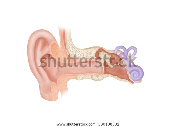
Ear Anatomy Without Labels Digital Art Stock Illustration 530108302
Picture of the Ear: Ear Conditions and Treatments - WebMD Earache: Pain in the ear can have many causes. Some of these are serious, some are not serious. Otitis media (middle ear inflammation): Inflammation or infection of the middle ear (behind the ...

30 Label Eye - Labels For Your Ideas
Ear Anatomy Images | McGovern Medical School Ear Anatomy Images. The ear drum is often transparent and looks like a stretched piece of clear plastic. The drum is approximately the size of a dime, with the newborn ear drum the same size as the adult. The malleus is the middle ear bone which is attached to the drum and easily identified. The middle ear space can be seen through the ear drum ...
Chap 10 Anatomy
TeachMeAnatomy - Making Anatomy Simple Containing over 1000 vibrant, full-colour images, TeachMeAnatomy is a comprehensive anatomy encyclopaedia presented in a visually-appealing, easy-to-read format.. Created by a team of doctors and medical students, each topic combines anatomical knowledge with high-yield clinical pearls, seamlessly bridging the gap between scholarly learning and improved patient care.
귀 해부 Ear Anatomy
heart diagram without labels Blank Ear Diagram | Human Ear Diagram, Ear Anatomy, Ear Diagram . ear diagram blank anatomy human eye drawing unlabeled worksheet parts label ears system quiz senses worksheets special physiology biology college. The Heart - Labelled Diagram wordwall.net. labelled ks4 gcse. DeLand Smiles: February 2012 delandsmiles.blogspot.com
Inner Ear Stock Images, Royalty-Free Images & Vectors | Shutterstock
Human Ear: Structure and Anatomy - Online Biology Notes Ear ossicles: The three ear ossicles (malleus, incus and stapes) form a chain of lever extending from tympanic membrane to inner ear. The ear ossicles transmit sound wave from ear drum to inner ear. Ear ossicles communicate the ear drum with internal ear through fenestra ovalis ( oval window). The ear ossicles are;
Post a Comment for "42 ear anatomy without labels"