38 diagram of the lungs with labels
Lungs label - Teaching resources Lungs label Examples from our community 4529 results for 'lungs label' Lungs Labelled diagram by Rbowerkail KS4 PE The Lungs Labelled diagram by Fayeroberts KS4 Y10 Biology Lungs Diagram Labelled diagram by Jon9 AC 4.1. Question 17 - Label the main components of the human lungs Labelled diagram by Phanvey494 The Lungs Labelled diagram Diagram Of The Respiratory System With Labels Stock Photos, Pictures ... In mammals and most other vertebrates, two lungs are located near the backbone on either side of the heart. Vector graphic. Lungs with Alveoli Labeled CG image of woman's chest area showing both lungs in isolation, with magnified view of alveoli air sacs labeled on faded flesh tone and white. Human Lungs
Lung Anatomy, Function, and Diagrams - Healthline The lungs begin at the bottom of your trachea (windpipe). The trachea is a tube that carries the air in and out of your lungs. Each lung has a tube called a bronchus that connects to the trachea....

Diagram of the lungs with labels
Human Throat Anatomy Pictures, Images and Stock Photos Human Respiratory System anatomical vector illustration, medical education cross section diagram with nasal cavity, throat, lungs and alveoli. Human Respiratory System anatomical vector illustration, medical education cross section diagram with nasal cavity, throat, esophagus, trachea, lungs and alveoli. human throat anatomy stock illustrations Lung Diagram | Free Lung Diagram Template - Edrawsoft The lung diagram template here clearly presents a pair of spongy on both side of the chest. Simply hitting on the template to learn more parts including pleura, ribs, bronchi, alveoli and more. Feel free to find out more human anatomy templates and symbols in the free download version. Download Template: Get EdrawMax Now! Free Download Popular Label the Lungs Diagram | Quizlet ... superior lobe of right lung ... middle lobe of right lung ... inferior lobe of right lung ... superior lobe of left lung ... left main (primary) bronchus ... lobar (secondary) bronchus ... segmental (tertiary) bronchus ... inferior lobe of left lung ... Sets found in the same folder Bi 233: Labeling the Larynx 21 terms SunshineGirl79 the cell
Diagram of the lungs with labels. The Lungs - Position - Structure - TeachMeAnatomy The lungs are roughly cone shaped, with an apex, base, three surfaces and three borders. The left lung is slightly smaller than the right - this is due to the presence of the heart. Apex - The blunt superior end of the lung. It projects upwards, above the level of the 1st rib and into the floor of the neck. Nervous System Worksheet Answers - WikiEducator 14/01/2008 · 8. The diagram below shows a section of a dog’s brain. Add the labels in the list below and, if you like, colour in the diagram as suggested. Cerebellum - blue; Spinal cord - green; Medulla oblongata - orange; Hypothalamus - purple; Pituitary gland - red; Cerebral hemispheres – yellow. 9. Match the descriptions below with the terms in the ... Label Lungs Diagram Printout - EnchantedLearning.com Read the definitions below, then label the lung anatomy diagram. bronchial tree - the system of airways within the lungs, which bring air from the trachea to the lung's tiny air sacs (alveoli). cardiac notch - the indentation in the left lung that provides room for the heart. diaphragm - a muscular membrane under the lungs. Lobes of the Lung - SmartDraw Lobes of the Lung Lobes of the Lung Create healthcare diagrams like this example called Lobes of the Lung in minutes with SmartDraw. SmartDraw includes 1000s of professional healthcare and anatomy chart templates that you can modify and make your own. 4/22 EXAMPLES EDIT THIS EXAMPLE Text in this Example: Lobes of the Lung
Circulatory System Diagram - Cardiovascular System and Blood ... They may come with or without labels. Common circulatory system diagrams show pulmonary circulation, coronary circulation, systematic circulation, veins, arteries, or a combination. The systemic circulation system is the most commonly illustrated of the systems that make up the circulatory system as it is the largest. Because the systemic circulation system is found in … Footprints-Science | GCSE science animations and quizzes | GCSE science … Biology random questions Cell structure Cell division Transport in cells Digestive system Heart and blood Health issues Plant tissues, organs and systems Communicable diseases Drugs Plant disease Photosynthesis Respiration Homeostasis Nervous system Hormones Reproduction Variation and Evolution Ecosystems Biodiversity Trophic levels Food production Chemistry … Acetylcholinesterase - Wikipedia Acetylcholinesterase (HGNC symbol ACHE; EC 3.1.1.7), also known as AChE, AChase or acetylhydrolase, is the primary cholinesterase in the body. It is an enzyme that catalyzes the breakdown of acetylcholine and some other choline esters that function as neurotransmitters.AChE is found at mainly neuromuscular junctions and in chemical synapses … labeled diagram of the lungs heart blood flow diagram lungs labeled arteries passes oxygenated major gets medicinebtg Cardiac Muscle. A. Longitud Inal Section Of Cardiac Muscle. Cells Are cardiac branched longitud inal cytoplasm striations nuclei Respiratory System Worksheet - WikiEducator
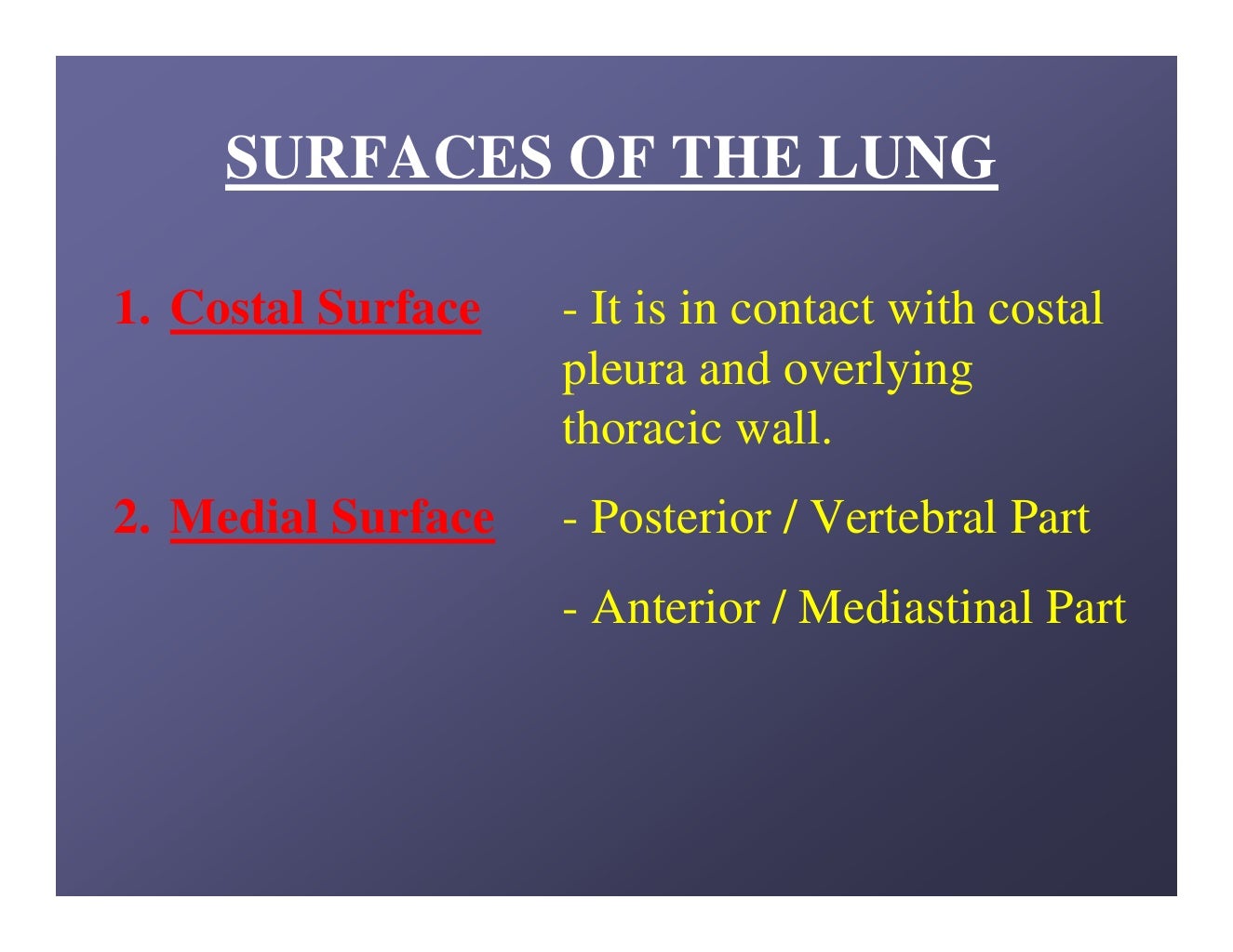
Heart Diagram with Labels and Detailed Explanation - BYJUS The diagram of heart is beneficial for Class 10 and 12 and is frequently asked in the examinations. A detailed explanation of the heart along with a well-labelled diagram is given for reference. Well-Labelled Diagram of Heart. The heart is made up of four chambers: The upper two chambers of the heart are called auricles. Lung diagram Images, Stock Photos & Vectors | Shutterstock Find Lung diagram stock images in HD and millions of other royalty-free stock photos, illustrations and vectors in the Shutterstock collection. Thousands of new, high-quality pictures added every day. Lungs: Anatomy, Function, and Treatment - Verywell Health They are the mediastinal surface, diaphragmatic surface, and costal surface. Lungs are protected by pleura, a thin layer of tissue that provides cushion and a small amount of fluid to help the lungs breathe smoothly. 1. Inside the lungs are bronchi—tubes that run from the trachea into each lung. The bronchi branch off into smaller tubes ... Labeled Diagram of the Human Lungs - Bodytomy Given below is a labeled diagram of the human lungs followed by a brief account of the different parts of the lungs and their functions. Each lung is enclosed inside a sac called pleura, which is a double-membrane structure formed by a smooth membrane called serous membrane.

Circulatory System - Birth Story
Human Heart (Anatomy): Diagram, Function, Chambers, Location … Pulmonary embolism: Typically a blood clot travels through the heart to the lungs. Heart valve disease : There are four heart valves, and each can develop problems. If …
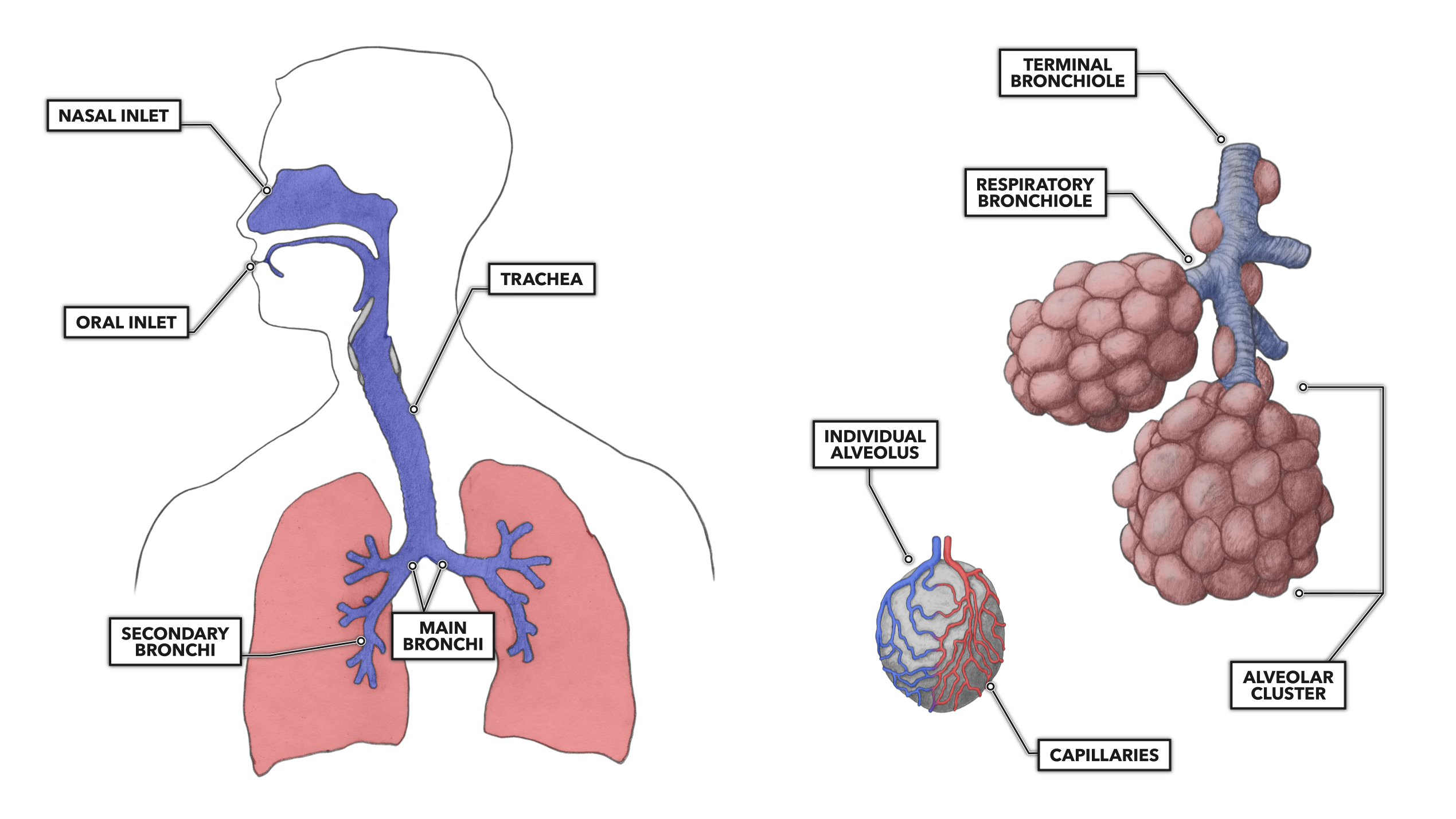
CrossFit | Lung Anatomy: The Airway and Alveoli
Human Respiratory System - Diagram - How It Works | Live Science During exhalation the diaphragm expands to force air out of the lungs. Adults normally take 12 to 20 breaths per minute. Strenuous exercise drives the breath rate up to an average of 45 breaths ...

SEPTUM AIR PURIFIER: Apr 16, 2010
The Lungs - Labelled diagram - Wordwall The Lungs - Labelled diagram trachea, bronchi, bronchioles, Alveoli, Heart, Diaphragm , Rib, Intercostal Muscle . The Lungs Share by Fayeroberts KS4 Y10 Biology Like Edit Content More Leaderboard Log in required Theme Switch template Interactives
Lung anatomy.
Respiratory System Anatomy, Diagram & Function | Healthline Respiratory. The respiratory system, which includes air passages, pulmonary vessels, the lungs, and breathing muscles, aids the body in the exchange of gases between the air and blood, and between ...

Circulation and respiration | Circulatory and respiratory systems | Siyavula
Lungs (Human Anatomy): Picture, Function, Definition, Conditions - WebMD The lungs are a pair of spongy, air-filled organs located on either side of the chest (thorax). The trachea (windpipe) conducts inhaled air into the lungs through its tubular branches, called...

Respitory system
Can you label the lungs? Quiz - PurposeGames.com Games by same creator. Back Muscles 3p Image Quiz. Labeling the Fetal Skull Bones. 7p Image Quiz. Labeling the Upper Leg Muscles 14p Image Quiz. Major Body Cavities 9p Image Quiz. Label the Pancreas 7p Image Quiz. Connective Tissues 10p Image Quiz. Can you Label the Heart 16p Image Quiz. 18p Image Quiz.
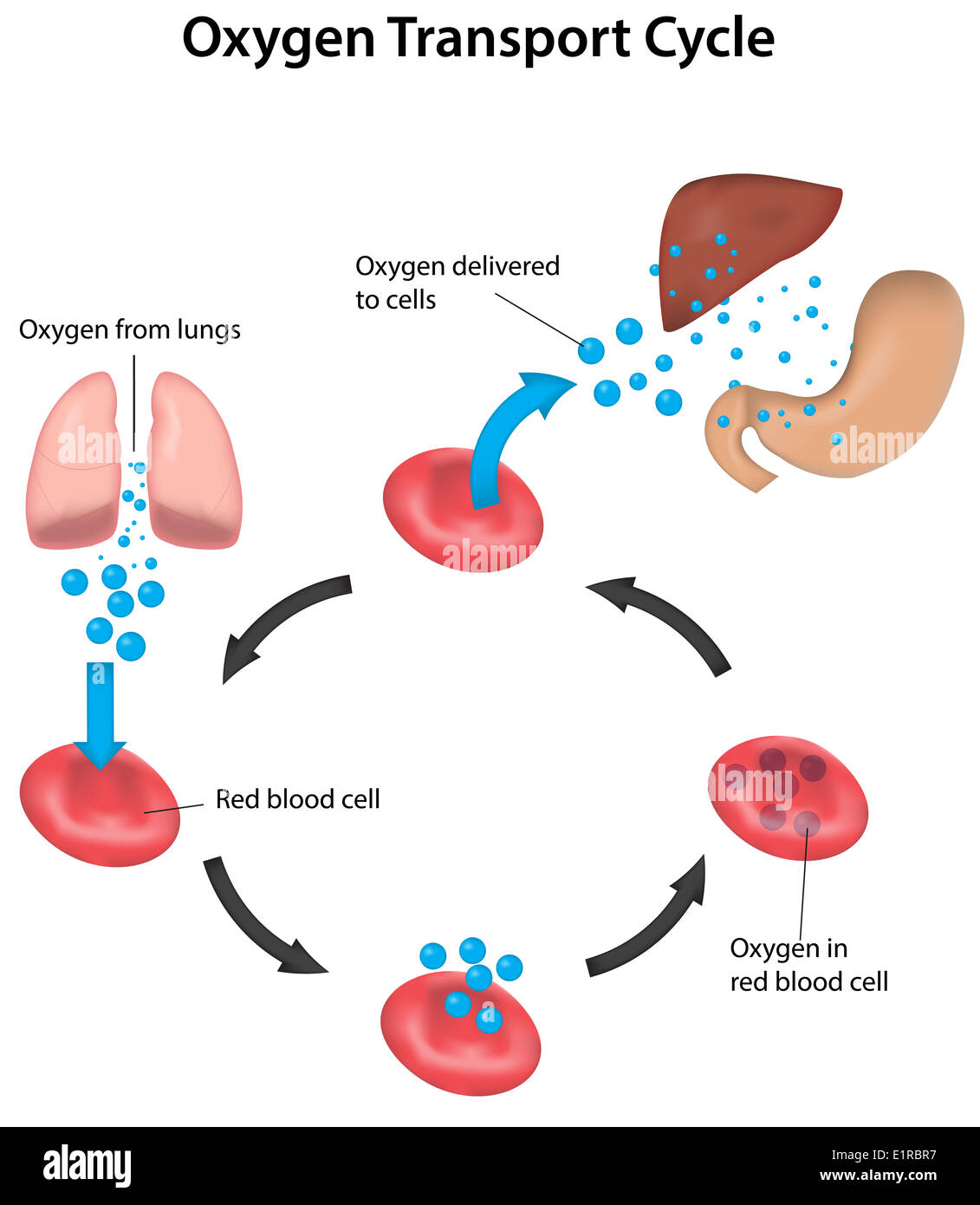
Oxygen Transport Cycle from Lungs to Organs Labeled Stock Photo - Alamy
Lung Diagram Photos and Premium High Res Pictures - Getty Images Browse 601 lung diagram stock photos and images available, or search for asthma or respiratory system to find more great stock photos and pictures. human lungs diagram - lung diagram stock illustrations. victorian anatomical drawing of human heart and lungs 19th century - lung diagram stock illustrations ...
Lung anatomy.
Female Anatomy Diagram High Resolution Stock Photography … Find the perfect female anatomy diagram stock photo. Huge collection, amazing choice, 100+ million high quality, affordable RF and RM images. No need to register, buy now!

181 best images about Respiratory system on Pinterest | Respiratory system, Smoking and Red ...
PDF ANATOMY OF LUNGS - University of Kentucky SURFACES OF THE LUNG 1. Costal Surface- It is in contact with costal pleura and overlying thoracic wall. 2. Medial Surface- Posterior / Vertebral Part - Anterior / Mediastinal Part Relations of Posterior Part 1. Vertebral Part 2. Intervertebral Discs 3. Posterior Intercostal Vessels 4. Splanchic Nerves RELATIONS OF ANTERIOR PART RIGHT SIDE 1.

CLASS BLOG: BIO 202 Respiratory System KEY
Fully Labelled Diagram Alveolus Lungs Showing Stock ... - Shutterstock Shutterstock customers love this asset! Stock Vector ID: 369984683 Fully labelled diagram of the alveolus in the lungs showing gaseous exchange. Vector Formats EPS 1114 × 800 pixels • 3.7 × 2.7 in • DPI 300 • JPG Vector Contributor S Steve Cymro Similar images Assets from the same collection Similar video clips

32 best Respiratory images on Pinterest | Anatomy, Anatomy reference and Lunges
respiratory diagram no labels blut zusammensetzung platelets labeled diagramm bloed. Digestive System Diagram En Clip Art At Clker.com - Vector Clip Art . system digestive diagram label clip digestion body clipart parts clker. Respitory System . respiratory respitory quizlet lungs. Respitory system.

White ramus communicans - wikidoc
Diagram Lungs Stock Illustrations - 2,543 Diagram Lungs ... - Dreamstime Download 2,543 Diagram Lungs Stock Illustrations, Vectors & Clipart for FREE or amazingly low rates! New users enjoy 60% OFF. 188,297,921 stock photos online. ... Internal vs external respiration system with air exchange outline diagram. Labeled educational breathing with oxygen in lungs and O2 in blood vessel from. Diaphragm anatomical vector ...
Lung Anatomy - EnchantedLearning.com
heart and lungs diagram labelled Arteries Of The Body Diagram — UNTPIKAPPS. . circulatory labeled arteries veins medicinebtg flow vessel artery systemic perfusion untpikapps cardiovascular capillaries exatin diagrams retinal segmentation fundus nerves arterial. Lungs anatomy human cavity thoracic respiratory labeled lung diagram label muscle system chest ...
Diagram of Air Tubes in the Lungs | ClipArt ETC
Lungs Diagram High Resolution Stock Photography and Images - Alamy This test is a rapid diag. ID: 2G0W7YJ (RF) Lung pulmonary alveoli or alveolus anatomy diagram as a medical concept of a close up of the human anatomy and respiratory or respiration medicine. ID: T1TJW8 (RF) Air Pollution and Lungs. ID: 2BEH12B (RM) Medical illustration of an asthmatic bronchiole in the human lungs.

Blog Sains Cikgu Kintan: 2010
The Respiratory System (Label Diagram) - ScienceQuiz.net Match each pair by dragging from right to left. When complete click Check button.

Learning Page: year 8 pass term 1 Body systems
Labeled diagram of the lungs/respiratory system. - SERC View Original Image at Full Size. Labeled diagram of the lungs/respiratory system. Image 37789 is a 1125 by 1408 pixel PNG Uploaded: Jan10 14. Last Modified: 2014-01-10 12:15:34

Lungs. Lungs Diagram. | Respiratory system, Lung anatomy, Respiratory system anatomy
Label the heart — Science Learning Hub 16/06/2017 · Drag and drop the text labels onto the boxes next to the heart diagram. If you want to redo an answer, click on the box and the answer will go back to the top so you can move it to another box. If you want to check your answers, use the Reset Incorrect button. This will reset incorrect answers only. When you are happy with your selection, use the Check answers …
Post a Comment for "38 diagram of the lungs with labels"